43 microscope diagram with labels and definitions
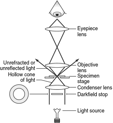
Gram Stain Technique - Amrita Vishwa Vidyapeetham Part 2: Labeling of the slides Drawing a circle on the underside of the slide using a glassware-marking pen may be helpful to clearly designate the area in which you will prepare the smear. You may also label the slide with the initials of the name of the organism on the edge of the slide. Diaphragm: Location, anatomy, innervation and function | Kenhub The diaphragm has two surfaces: thoracic and abdominal. The thoracic diaphragm is in contacts with the serous membranes of the heart and lungs; namely, the pericardium and pleura. The abdominal diaphragm is in direct contact with the liver, stomach, and spleen. Abdominal surface of the diaphragm (diagram)
Medicine & Health | UNSW Sydney Research & impact. UNSW Medicine & Health is world renowned for its research and impact addressing issues of health disparities and improving lives through our key research themes of Cancer; Infectious Disease, Immunity & Inflammation; Neuroscience, Mental Health & Addiction; Cardiac, Vascular & Metabolic Medicine and Health Systems.

Microscope diagram with labels and definitions
WHMIS 2015 - Pictograms : OSH Answers - Canadian Centre for ... Suppliers and employers must use and follow the WHMIS 2015 requirements for labels and safety data sheets (SDSs) for hazardous products sold, distributed, or imported into Canada. Please refer to the following OSH Answers documents for information about WHMIS 2015: WHMIS 2015 - General. WHMIS 2015 - Labels. scheme work biology - Free KCPE Past Papers Definition . Use of magnifying lens. By the end of the lesson, the learner should be able to: ... Draw and label the light microscope; Description of a cell; Drawing and labeling the light microscope . Light microscope; Diagram of light microscope; Comprehensive secondary Biology students Bk. 1 page 17; Teachers bk. 1 pages 11-19; DP Biology: Calculating Magnification and Size Activity 1 Calculating magnification of an image using it's scale bar The three images below (click the eye to reveal) show a worked example of how to calculate sizes of cells organelles from electron micrographs step by step. Follow these steps carefully then complete the calculations on the worksheet.
Microscope diagram with labels and definitions. LabVIEW - NI Community Discuss developing automated research, validation, and production test systems in NI's graphical programming environment. Product Documentation. • NI Product Documentation Center. • Release Notes. • Knowledge Base. NI Learning Center. • Getting Started. • Introduction to LabVIEW. • LabVIEW Core 1. Spinal Cord Cross Section | New Health Advisor The tracks that move upward are responsible for signals to the brain and the descending tracts send the signals from the brain to neurons throughout the body. 2. Gray Matter In the center of the gray matter you will find the cerebrospinal fluid. The specific horns of the gray matter are responsible for different things. Fluorescence In Situ Hybridization (FISH) - Genome.gov In this technique, the full set of chromosomes from an individual is affixed to a glass slide and then exposed to a "probe"—a small piece of purified DNA tagged with a fluorescent dye. The fluorescently labeled probe finds and then binds to its matching sequence within the set of chromosomes. Biology Archive | October 06, 2022 | Chegg.com correct the answers/blanks. The smallest functional unit of a skeletal muscle is the and when the muscle contracts the changes in length. The nervous system is subdivided into the nervous system and the system, which receives input from the nervous system. At low temperatures the coagulation is often less efficient.
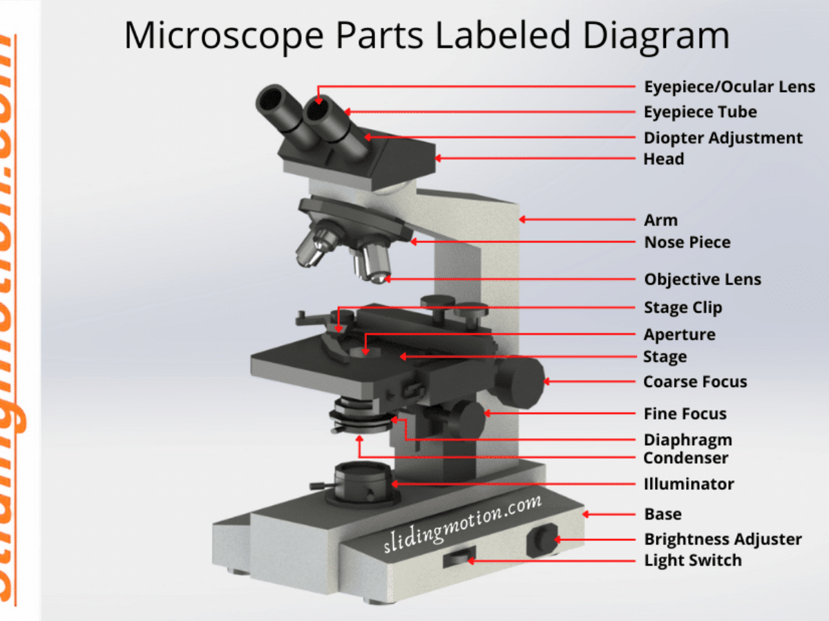
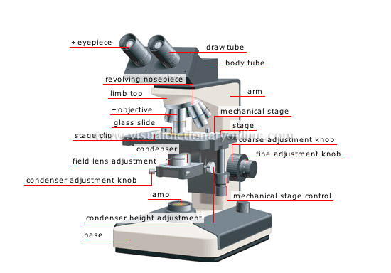
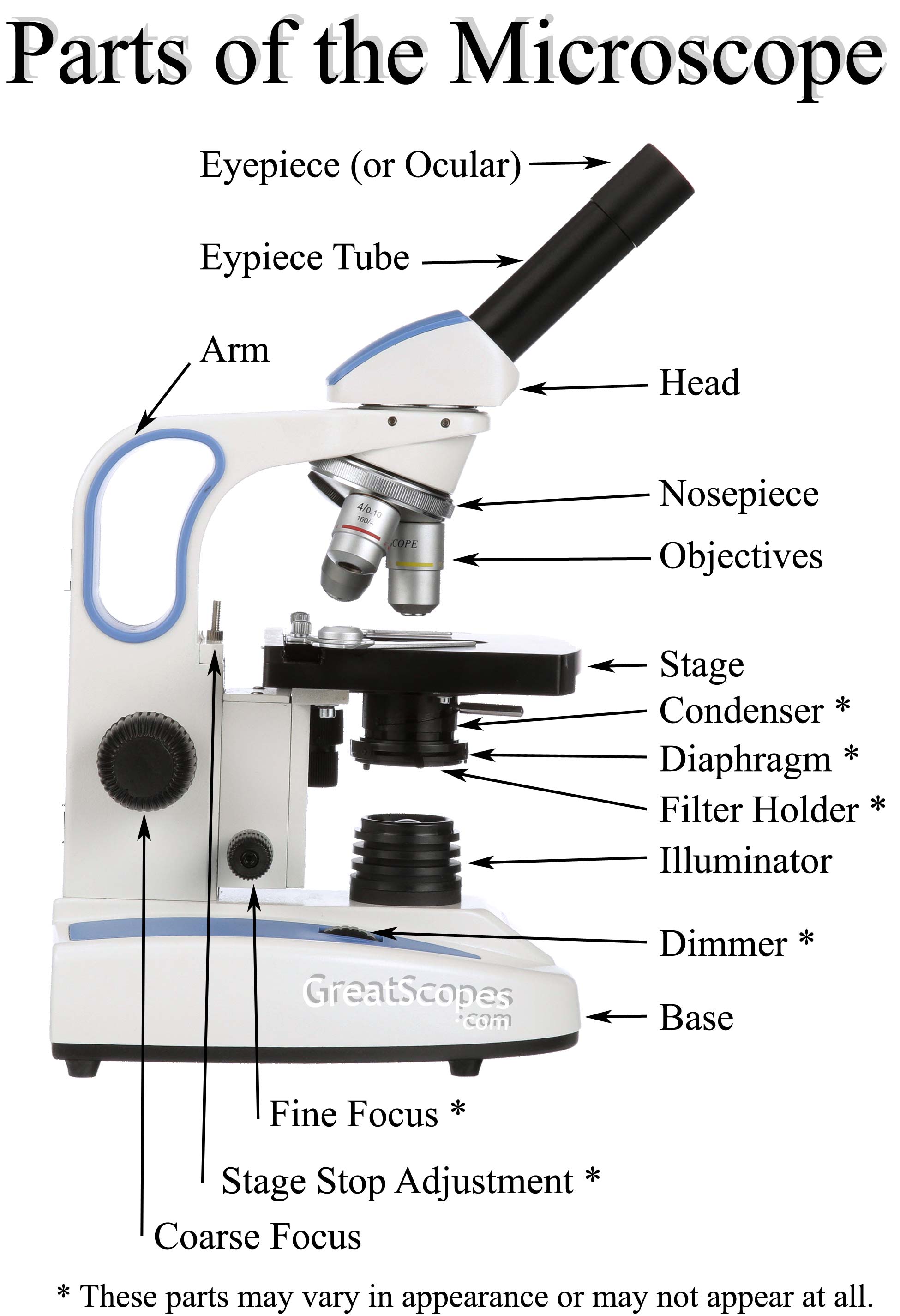
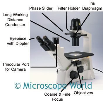
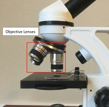
Binocular Microscope Anatomy - Parts and Functions with a Labeled Diagram The important non-optical parts of the light compound microscope are the body tube or head, arm or frame, fine adjustment, coarse adjustment, nose piece, stage, and base. Now, I will describe all these non-optical parts of the light compound microscope with the labeled diagrams. The body tube of the microscope Mineralogy Database Complete, up-to-date, mineral database containing 4,714 mineral species descriptions and comprehensive picture library of images. These data are linked to mineral tables by crystallography, chemical composition, physical and optical properties, Dana classification, Strunz classification, mineral name origins, mineral locality information, and alphabetical listing of all known valid mineral ... X-Ray Generation Notes - University of Oklahoma X-Ray photons are electromagnetic radiation with wavelengths typically in the range 0.1 - 100 Å. X Rays used in diffraction experiments have wavelengths of 0.5 - 1.8 Å. X Rays can be produced by conventional generators, by synchrotrons, and by plasma sources. Electromagnetic radiation from nuclear reactions, called γ radiation, can also ... Home Lab Beginners guide - Hardware - Hayden James A rack unit (abbreviated U or RU) is a unit of measure defined as 1 3⁄4 inches (or 44.45 mm). It's the unit of measurement for the height of 19-inch and 23-inch rack frames and the equipment's height. The height of the frame/equipment is expressed as multiples of rack units. A typical full-size rack is 42U high.
X-ray crystallography - Wikipedia X-ray crystallography is the experimental science determining the atomic and molecular structure of a crystal, in which the crystalline structure causes a beam of incident X-rays to diffract into many specific directions. By measuring the angles and intensities of these diffracted beams, a crystallographer can produce a three-dimensional picture of the density of electrons within the crystal. Light Microscope (Theory) - Amrita Vishwa Vidyapeetham Parts of a Microscope It consists of mainly three parts: Mechanical part - base, c-shaped arm and stage. Magnifying part - objective lens and ocular lens. Illuminating part - sub stage condenser, iris diaphragm, light source. Mechanical part Base: It helps in holding the various parts of microscope. It also contains the light source. Oxygen Saturation (02 Sat): Normal Ranges and How to Raise It An ABG value refers to the levels of oxygen and carbon dioxide in blood running through your veins. During an ABG, a nurse or lab technician draws blood from an artery, such as the radial artery in the wrist or the femoral artery in the groin. The sample is immediately analyzed by a machine or in a lab. Colony Morphology of Bacteria - Microbe Online Bacteria grow on solid media as colonies. A colony is defined as a visible mass of microorganisms originating from a single mother cell. Key features of these bacterial colonies serve as important criteria for their identification. Characteristics of bacterial colonies Colony morphology can sometimes be useful in bacterial identification.
Let's Learn! What are Microscopes? : Olympus Kids Class ...
leaf | Definition, Parts, & Function | Britannica stomate, also called stoma, plural stomata or stomas, any of the microscopic openings or pores in the epidermis of leaves and young stems. Stomata are generally more numerous on the underside of leaves. They provide for the exchange of gases between the outside air and the branched system of interconnecting air canals within the leaf.
Microscope | definition of microscope by Medical dictionary
Electrical Safety - Basic Information : OSH Answers Label all circuit breakers and fuse boxes clearly. Each switch should be positively identified as to which outlet or appliance it is for. Do not use outlets or cords that have exposed wiring. Do not use portable cord-and-plug connected power tools if the guards are removed. Do not block access to panels and circuit breakers or fuse boxes.

This is a common compound microscope. Label its parts from A ...
StudiousGuy - Your Study Buddy Types of Hire Purchase. October 6, 2022 Commerce. Black Power Movement
16 Basic Parts of Microscope, Function, Names & Labeled Diagram
Mr. Jones's Science Class Earth, Moon, & Sun System (PPT.) Seasons Interactive (Online Activity) Moon Phases - Introductory Activity. Modeling the Phases of the Moon. Problems in Space (Online Activity) Lunar & Solar Eclipses - Webquest.

Light Microscope Parts, Function & Uses | What is a Light Microscope? Video
Electron Microscope: Principle, Types, Applications Schematic diagram of Transmission Electron Microscope Like any ordinary microscope, the electron microscope also uses a light source, a combination of lenses to produce a magnified image, however, this vary slightly as compared to ordinary light microscope. Source of light: The source of light is replaced by a beam of very fast-moving electrons.

Label the microscope — Science Learning Hub
Micro Definition & Meaning - Merriam-Webster The meaning of MICRO is very small; especially : microscopic. Recent Examples on the Web: Adjective No, the micro-mini isn't going away, but runways from the likes of Altuzarra and Tory Burch showed that floor-skimming maxi skirts will be everywhere come spring. — Dale Arden Chong, ELLE, 4 Oct. 2022 Kevin Kruse is the Founder + CEO of LEADx, a leadership development system that scales and ...

Compound Microscope Parts, Function, & Diagram | What is a ...
Cell Division: Mitosis and Meiosis - Owlcation Chromosome - A molecule of DNA wrapped around histones that becomes visible during prophase of cell division. Chromatid - A replicated chromosome: each strand of the 'X' is a chromatid. Diploid - Cells that have two copies of each chromosome in their nuclei. Haploid - Cells that have one copy of each chromosome in their nuclei.

Microscope World Blog: What is a Compound Microscope?
Diagram of Human Heart and Blood Circulation in It Four Chambers of the Heart and Blood Circulation. The shape of the human heart is like an upside-down pear, weighing between 7-15 ounces, and is little larger than the size of the fist. It is located between the lungs, in the middle of the chest, behind and slightly to the left of the breast bone. The heart, one of the most significant organs ...
The Compound Light Microscope
Quantum dot - Wikipedia Quantum dots (QDs) are semiconductor particles a few nanometres in size, having optical and electronic properties that differ from those of larger particles as a result of quantum mechanics.They are a central topic in nanotechnology.When the quantum dots are illuminated by UV light, an electron in the quantum dot can be excited to a state of higher energy.
SCIENCE :: PHYSICS: OPTICS :: MAGNIFYING GLASS AND ...
Microscope: Types of Microscope, Parts, Uses, Diagram - Embibe Microscope: Definition, Parts, Parameters, Types, & Uses A microscope is an instrument that produces enlarged images of small objects. It provides the observer an exceedingly close view of minute structures at a scale convenient for examination and analysis. The microscope magnifies microscopic objects that are not visible to the naked eyes.

Microscope labeled diagram
Metaphase - Genome.gov During metaphase, the nucleus dissolves and the cell's chromosomes condense and move together, aligning in the center of the dividing cell. At this stage, the chromosomes are distinguishable when viewed through a microscope. Metaphase chromosomes are used in karyotyping, a laboratory technique for identifying chromosomal abnormalities. Narration
Glossary :: GreatScopes
DP Biology: Calculating Magnification and Size Activity 1 Calculating magnification of an image using it's scale bar The three images below (click the eye to reveal) show a worked example of how to calculate sizes of cells organelles from electron micrographs step by step. Follow these steps carefully then complete the calculations on the worksheet.

microscope | Types, Parts, History, Diagram, & Facts | Britannica
scheme work biology - Free KCPE Past Papers Definition . Use of magnifying lens. By the end of the lesson, the learner should be able to: ... Draw and label the light microscope; Description of a cell; Drawing and labeling the light microscope . Light microscope; Diagram of light microscope; Comprehensive secondary Biology students Bk. 1 page 17; Teachers bk. 1 pages 11-19;

label microscope diagram | Charts | Microscope, Anatomy bones ...
WHMIS 2015 - Pictograms : OSH Answers - Canadian Centre for ... Suppliers and employers must use and follow the WHMIS 2015 requirements for labels and safety data sheets (SDSs) for hazardous products sold, distributed, or imported into Canada. Please refer to the following OSH Answers documents for information about WHMIS 2015: WHMIS 2015 - General. WHMIS 2015 - Labels.
microscope - Kids | Britannica Kids | Homework Help
Parts of a Microscope | Microscope Parts and Functions | Labkafe
Anatomy of a Microscope | Microscopy Primer | Olympus LS

Compound Microscope Parts – Labeled Diagram and their ...

Microscope Diagram Labeled, Unlabeled and Blank | Parts of a ...

A Study of the Microscope and its Functions With a Labeled ...
Microscope World Blog: Inverted Biological Microscopes
Parts of a microscope with functions and labeled diagram

Labeling the Parts of the Microscope | Microscope World Resources

Biology Microscope Labeling and Definitions (Light/Compound ...

Types of Microscopes: Definition, Working Principle, Diagram ...

Electron microscope - Wikipedia

Parts of a Compound Microscope and Their Functions

Parts of a Microscope Diagram | Quizlet

Parts of a microscope with functions and labeled diagram

Dissecting Stereo Microscope Parts and Functions
What Is the Function of a Microscope?

Compound Microscope: Know Definition,working, diagram, properties

Simple Microscope - Parts, Functions, Diagram and Labelling ...

Compound Microscope: Definition, Parts, Application, Working ...

Compound Microscope- Definition, Labeled Diagram, Principle ...

Simple Microscope - Diagram (Parts labelled), Principle ...

Lesson Explainer: Microscopy | Nagwa

Microscope Parts | Microbus Microscope Educational Website
microscope | Types, Parts, History, Diagram, & Facts | Britannica

Compound Microscope Parts, Functions, and Labeled Diagram ...

The Microscope
16 Parts of a Compound Microscope: Diagrams and Video ...

What is a Compound Microscope? | Microscope World Blog
Post a Comment for "43 microscope diagram with labels and definitions"